
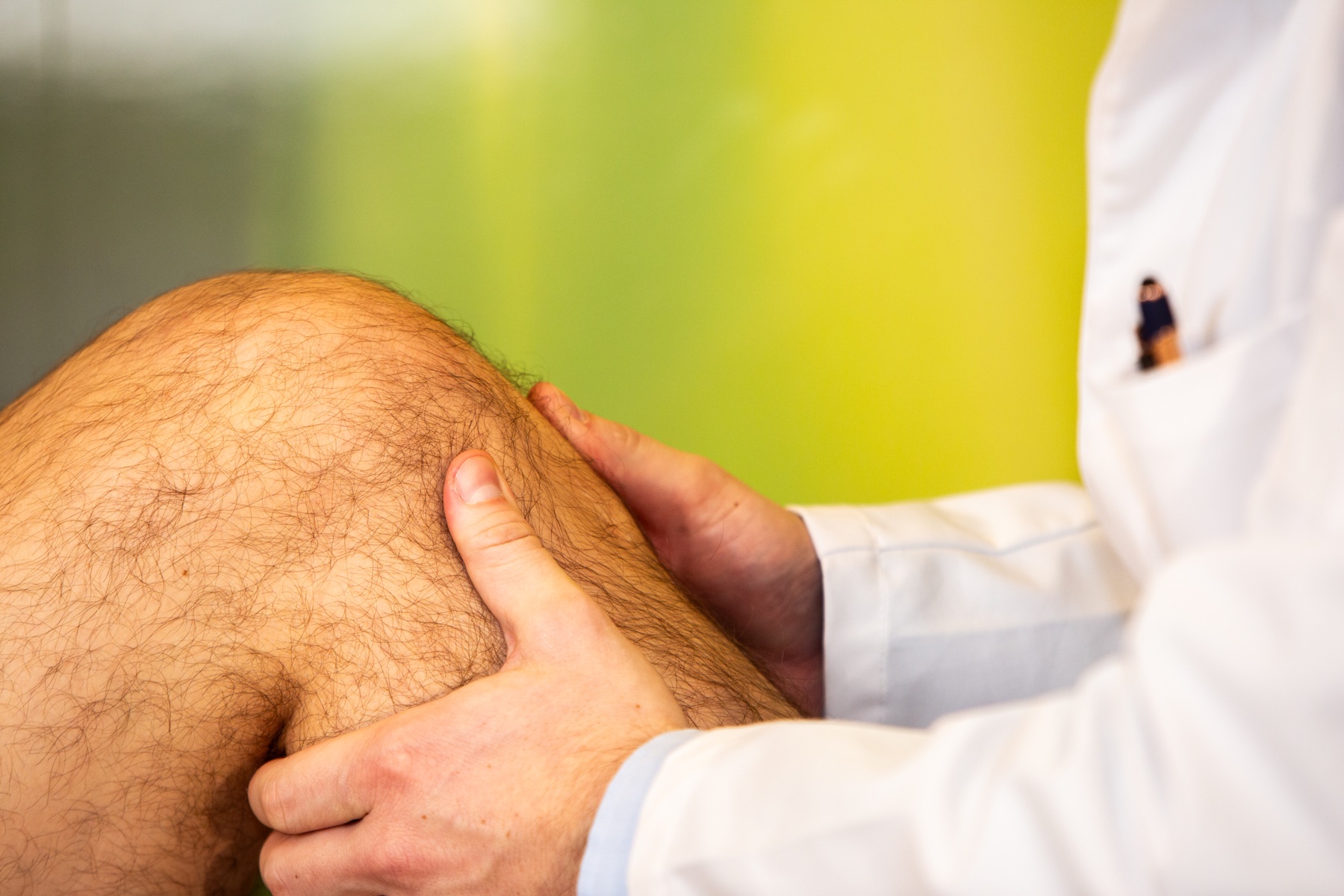
Not every meniscus tear needs surgery. The decision depends not only on the MRI findings or the size of the tear, but on its pattern, stability, symptoms, mobility, cartilage condition, associated injuries and the individual's activity goals.
For many stable and especially degenerative meniscal lesions, structured non-surgical treatment is the first step. By contrast, early meniscus-preserving surgery may be appropriate for a displaced fragment causing true locking, certain repairable traumatic tears or a suitable meniscal root tear.
Biological treatments are now also available between physiotherapy and surgery, including platelet-rich plasma (PRP) made from the patient's own blood and, for selected degenerative findings, Microfat or microfragmented adipose tissue.
Key points
- A meniscus tear on MRI does not automatically mean that surgery is needed.
- For many stable degenerative tears without true locking, active, structured rehabilitation is initially the most important treatment.
- An acutely locked knee, a displaced repairable fragment, or certain root tears and larger radial tears should be assessed by a specialist early.
- If surgery is necessary, as much functional meniscal tissue as possible should be preserved.
- PRP may be a biological treatment option for selected stable meniscal lesions. Clinical improvements have been described; however, structural healing of the meniscus cannot be guaranteed.
- Microfat may be discussed for selected combinations of degenerative meniscal changes, cartilage damage and osteoarthritis. The meniscus-specific evidence is currently less extensive than for PRP.
- A second opinion is particularly useful if partial meniscectomy has been recommended, or if it is unclear whether the MRI findings actually explain the symptoms.
A brief explanation: what is a meniscus tear?
There are two menisci in the knee: the medial meniscus on the inner side and the lateral meniscus on the outer side. They consist of resilient fibrocartilage and distribute forces between the thigh bone and shin bone.
The menisci are therefore much more than simple “shock absorbers”. They contribute to load distribution, joint stability, joint lubrication and protection of the articular cartilage.
A meniscus tear can result from an acute injury, for example a twisting movement during sport. More often, however, meniscal changes develop gradually as the tissue structure changes.
This distinction matters for treatment.
An MRI finding does not yet answer the treatment question
A common sequence is:
Knee pain → MRI → “meniscus tear” → recommendation for surgery.
The decision should not be made that simply.
Meniscal changes are common even in people without knee pain, especially in middle and older age. The first question to clarify is therefore:
Does this particular tear actually match the patient's symptoms?
Relevant factors include:
- injury mechanism
- location of pain
- joint effusion
- mobility
- true locking
- knee stability
- cartilage damage
- osteoarthritis
- leg alignment
- sporting demands
- previous treatment.
The written MRI report does not always fully describe the tear pattern either.
For a thorough assessment, the original MRI images should therefore be reviewed whenever possible, not just the radiology report.
What types of meniscus tear are there?
The tear pattern has a major influence on treatment.
Degenerative meniscus tear
Degenerative meniscus tears occur in structurally altered tissue and are often associated with the following:
- cartilage wear
- meniscal extrusion
- knee osteoarthritis
- subchondral changes
These associated changes are common.
A slight twisting force can trigger symptoms for the first time, even though the meniscal change was already present.
For stable degenerative lesions without true locking, structured non-surgical treatment is usually the appropriate first step.
Traumatic meniscus tear
A traumatic tear occurs in connection with a specific injury, for example:
- while playing football
- while skiing
- during rapid changes of direction
- through a twisting movement under load.
Here too, “traumatic” does not automatically mean “surgery”.
Non-surgical treatment may also be reasonable for a stable isolated tear without locking.
However, one question is particularly important:
Is the tear repairable, and could waiting lead to the loss of valuable meniscal function?
Bucket-handle tear and displaced fragment
In a bucket-handle tear, an elongated fragment of the meniscus can become displaced towards the centre of the joint.
If this suddenly prevents full straightening of the knee, true mechanical locking is suspected.
This should be assessed promptly by an orthopaedic specialist.
The key question is not:
“How much meniscus do we need to remove?”
Instead, the first question is:
“Can the meniscus be repositioned and preserved?”
Radial tear
A radial tear runs across the circular fibres of the meniscus.
Larger complete radial tears can substantially impair the meniscus's ability to transmit load through circumferential tension, known as hoop stress.
These findings must be assessed differently from small, stable degenerative changes.
Meniscal root tear
The meniscal roots anchor the meniscus to the top of the shin bone.
A complete root tear can substantially reduce the meniscus's ability to transmit load and cause it to move progressively out of the joint space.
Meniscal root repair may therefore be appropriate for suitable patients without advanced osteoarthritis.
The degree of osteoarthritis, cartilage condition, meniscal extrusion and leg alignment must be included in this decision.
Horizontal tear
Horizontal tears often occur in degeneratively altered meniscal tissue.
They may be symptom-free, but can also cause activity-related pain along the joint line.
Stable horizontal lesions without locking are among the findings for which non-surgical and, where appropriate, biological treatments can be discussed initially.
Cracking is not the same as a locked knee
Many patients report:
- cracking
- clicking
- occasional catching
- grinding
- snapping.
These symptoms alone are not a reliable indication for surgery.
True mechanical locking means that a displaced tissue fragment physically prevents movement.
This must be distinguished from:
- pain-limited straightening
- a protective posture because of joint effusion
- temporary stiffness
- cracking without loss of movement.
This distinction is extremely important clinically.
How is a meniscus tear properly diagnosed?
Medical history
The initial questions are:
- Was there an injury?
- Where is the pain?
- When does it occur?
- Does the knee swell after activity?
- Can the knee be fully straightened?
- Does the knee feel unstable?
- Which sports does the patient take part in?
Clinical examination
The examination includes:
- mobility
- tenderness along the joint line
- joint effusion
- stability
- meniscus-specific provocation tests
- leg alignment
- functional loading.
MRI
MRI is the most important non-invasive imaging method for assessing the meniscus.
In addition to the meniscus, the MRI assessment includes:
- cartilage
- cruciate ligaments
- collateral ligaments
- bone
- joint effusion
- synovial membrane
- patellofemoral joint
These structures and findings are assessed together on MRI.
X-rays
Weight-bearing X-rays may be crucial when symptoms are degenerative.
A meniscus tear in an otherwise largely healthy knee requires a different treatment assessment from the same MRI label in a knee with established, pronounced osteoarthritis.
Meniscus tear: when is surgery appropriate?
Surgery should be considered particularly for:
- true mechanical locking that prevents straightening because of a displaced fragment
- a repairable, unstable traumatic tear
- certain bucket-handle tears
- a suitable meniscal root tear
- a large radial tear that significantly affects function
- a tear combined with a ligament injury requiring surgery
- repeated, objectively demonstrable catching with a corresponding unstable fragment
- significant symptoms despite well-delivered non-surgical treatment, when the findings and symptoms clearly match.
Important: even when surgery is performed, the aim today is not to remove as much meniscus as possible, but to preserve as much functional meniscal tissue as possible.
When is surgery usually not the first step?
Immediate partial meniscectomy is often not the initial treatment for:
- a degenerative meniscus tear without locking
- a stable tear
- an incidental MRI finding
- non-specific cracking
- knee osteoarthritis as the main problem
- pronounced cartilage damage
- a knee that has not yet undergone structured rehabilitation.
Particularly for degenerative meniscal lesions, high-quality randomised studies show that structured physiotherapy can achieve long-term functional outcomes comparable to early arthroscopic partial meniscectomy.
Meniscal repair or partial meniscectomy?
Meniscal suturing and repair
If a tear is repairable, the aim is to stabilise the meniscus and allow it to heal biologically.
This is particularly relevant for:
- suitable longitudinal tears
- bucket-handle tears
- selected radial tears
- root injuries.
The main advantage is:
Meniscal tissue, and therefore meniscal function, is preserved.
However, rehabilitation takes longer than after partial resection.
Limited partial meniscectomy
If an unstable part of the meniscus cannot reasonably be repaired from a biological and mechanical perspective, limited partial resection may be necessary.
Only as much tissue as necessary should be removed.
The greater the loss of functional meniscal tissue, the more the distribution of load in the knee changes over the long term.
Partial meniscectomy should therefore not be regarded as an equivalent substitute for a meniscal repair that is technically and biologically appropriate.
What does good non-surgical treatment involve?
“Non-surgical” does not mean simply resting the knee for several weeks.
Depending on the findings, structured treatment includes:
- temporarily adjusting activities and loads that trigger pain
- improving mobility
- quadriceps training
- hamstring training
- hip and trunk stability
- neuromuscular training
- improving control of leg alignment
- balance training
- progressively rebuilding tolerance to running and jumping
- sport-specific rehabilitation
- treatment of joint effusion
- treatment of associated osteoarthritis
- individual management of activity and loading.
Rehabilitation should be guided by the knee's response, not only by calendar weeks.
Can PRP be useful for a meniscus tear?
Yes. PRP may be discussed as a biological treatment option for selected stable meniscal lesions that do not cause locking.
PRP stands for platelet-rich plasma.
The patient's own blood is collected and processed to obtain a concentrated fraction of platelets.
Platelets contain numerous bioactive signalling substances that can influence repair and inflammatory processes.
However, PRP is not a uniform product. Differences include:
- platelet concentration
- white blood cell content
- number of injections
- injection site
- preparation method.
What does the research show?
Studies of non-surgical PRP treatment for meniscal lesions show improvements in pain and function in selected patients.
Imaging results are more variable: some studies have described changes or signs of healing on MRI, but structural healing can by no means be guaranteed.
Recent systematic reviews currently identify PRP as the most extensively clinically studied injection therapy for meniscal lesions. At the same time, differences between studies and the PRP protocols used remain relevant.
PRP may be discussed particularly for:
- stable degenerative lesions
- certain horizontal tears
- persistent symptoms despite structured rehabilitation
- associated cartilage irritation
- early or moderate knee osteoarthritis.
When a displaced fragment causes mechanical locking, PRP cannot replace necessary mechanical stabilisation.
What is the role of PRP when knee osteoarthritis is also present?
Degenerative meniscal changes often do not occur in isolation.
They are frequently accompanied by:
- cartilage changes
- synovial irritation
- meniscal extrusion
- early or moderate osteoarthritis.
PRP has now been investigated for knee osteoarthritis in a large number of clinical and randomised studies.
The work of Boffa and colleagues shows, in particular, that the scientific evidence base for PRP has become considerably more extensive than some older guideline assessments might suggest.
However, this does not mean that PRP is the right treatment for every patient.
The decisive factors remain:
- degree of osteoarthritis
- tear pattern
- joint effusion
- leg alignment
- age
- activity level
- treatment goal.
Is Microfat an alternative for a meniscus tear?
For selected combinations of degenerative findings, Microfat may be discussed as a cell-based biological treatment.
Microfat, or microfragmented adipose tissue, is obtained from the patient's own fat tissue and processed mechanically.
The tissue contains, among other components:
- stromal cell populations
- perivascular cell populations
- extracellular matrix
- numerous biological signalling structures.
Microfat is therefore often referred to informally as “stem cell therapy”.
A more medically precise description is:
cell-based regenerative therapy using minimally manipulated autologous adipose tissue.
In which situation is Microfat of particular interest?
The situation of greatest clinical interest at present is not an isolated meniscus tear causing acute locking, but a degeneratively altered knee with a combination of:
- meniscal degeneration
- cartilage damage
- synovial irritation
- early or moderate osteoarthritis.
Meniscus-specific studies show interesting clinical signals, but the evidence base is substantially smaller than for PRP.
Randomised studies in knee osteoarthritis also show clinical improvements after both Microfat and PRP, without establishing general superiority of Microfat.
Microfat should therefore not be seen as “better PRP”, but as a biologically different procedure with its own indications.
Physiotherapy, PRP, Microfat or surgery?
| Finding | Often appropriate initial approach |
|---|---|
| Stable degenerative tear without locking | Structured rehabilitation |
| Stable tear with symptoms despite good rehabilitation | Reassess the diagnosis; PRP may be discussed |
| Combined degenerative meniscal changes, cartilage damage and osteoarthritis | Osteoarthritis-focused treatment; discuss PRP and, in selected cases, Microfat |
| Repairable traumatic tear | Weigh meniscus-preserving surgery against a non-surgical strategy |
| Bucket-handle tear with true locking | Promptly assess the possibility of surgical repositioning and suturing |
| Suitable meniscal root tear | Assess the possibility of repair early |
| Large complete radial tear | Assess its functional significance and repairability |
| Advanced osteoarthritis | Do not automatically treat the meniscus; assess the overall knee disease |
This overview does not replace an individual treatment decision.
A perspective from clinical practice
“With a meniscus tear, the MRI label should not determine the treatment. What matters is whether the tear pattern, clinical findings, symptoms and activity goals really fit together. Particularly before a planned partial meniscectomy, it is worth asking whether the meniscus can be preserved, or whether a non-surgical strategy is medically equivalent or more appropriate.”
PD Dr. med. Daniel P. Berthold, specialist in orthopaedics and trauma surgery
Second opinion before meniscus surgery: what should be checked?
A good second opinion answers more than:
“Surgery: yes or no?”
It should clarify:
1. Is the meniscus actually causing the symptoms?
The location of pain, pattern of activity-related symptoms and examination findings must match the MRI.
2. What is the tear pattern?
Particularly relevant features include:
- tear direction
- tear length
- stability
- displacement
- blood supply zone
- proximity to the root
- meniscal extrusion.
3. What is the condition of the rest of the knee?
The assessment must also include:
- cartilage
- cruciate ligaments
- collateral ligaments
- bone
- patellofemoral joint
- leg alignment.
4. Can the meniscus be preserved?
When deciding on surgery, the following should be clarified beforehand:
- Is meniscal suturing possible?
- Is meniscal root repair necessary?
- Under what circumstances would partial resection be performed during the operation?
5. Has non-surgical treatment really been adequate?
The number of physiotherapy appointments is not the deciding factor.
What matters is:
- exercise content
- the amount of loading
- progression
- adherence
- the knee's response.
6. Which biological option is appropriate?
For stable lesions, PRP or, in more complex degenerative changes, Microfat may also be discussed depending on the findings.
What should I bring to a second-opinion appointment?
Ideally, bring:
- original MRI images or DICOM data
- the radiology MRI report
- any existing X-rays
- previous MRI examinations
- operation reports from earlier procedures
- a summary of treatment to date
- information about the operation currently being proposed.
It is particularly important to know:
Is meniscal suturing, root repair or partial meniscectomy planned?
Questions to ask before meniscus surgery
- Which finding is most likely to be causing my symptoms?
- Is the knee truly mechanically locked?
- What is the exact tear pattern?
- Can the meniscus be sutured?
- If partial resection is recommended, why?
- How much meniscal tissue would need to be removed?
- Which non-surgical options have already been adequately tried?
- Is PRP a reasonable option for my findings?
- Is there also significant cartilage damage or osteoarthritis?
- How does rehabilitation differ after suturing and partial resection?
- When is a return to my sport realistic?
- What is plan B if the chosen treatment does not help enough?
When can I return to sport with a meniscus tear?
Return to sport should not be based solely on a fixed number of weeks.
Relevant factors include:
- a largely settled knee without significant irritation
- no significant swelling in response to activity
- unrestricted mobility or sufficient mobility for the sport
- restored muscle strength
- good control of leg alignment
- safe single-leg loading
- sport-specific activity tolerance.
After meniscal suturing, rehabilitation is specific to the tear and operation and is generally considerably more cautious than after partial resection.
For a meniscus tear treated without surgery, loading is increased gradually according to the knee's response.
Conclusion: meniscal function matters, not the MRI label
A meniscus tear is not a single uniform diagnosis.
For many degenerative and stable lesions without locking, structured non-surgical treatment is the right first step.
By contrast, meniscus-preserving surgery may be appropriate for certain unstable, displaced, root-adjacent or functionally important tears.
PRP extends the range of non-surgical treatments, particularly for selected stable lesions and combinations of degenerative meniscal and cartilage changes. Microfat provides another biological option for selected degeneratively altered knees, although its evidence base is smaller and the indication should be established particularly carefully.
If surgery has already been recommended, the key question should therefore not be:
“How quickly can the meniscus be operated on?”
Instead, the question is:
“For these particular findings, which treatment preserves as much knee function as possible over the long term?”
Second opinion for a meniscus tear in Munich
An orthopaedic second opinion assesses the clinical findings, original MRI images, tear pattern, cartilage condition, associated injuries and previous treatment together.
The aim is an understandable medical decision, whether the eventual treatment is non-surgical, biological or surgical.
Medical information
This information does not replace an in-person medical examination.
If the knee suddenly cannot be straightened, there is pronounced swelling after an injury, an inability to bear weight, marked warmth, redness or fever, prompt medical assessment is needed.